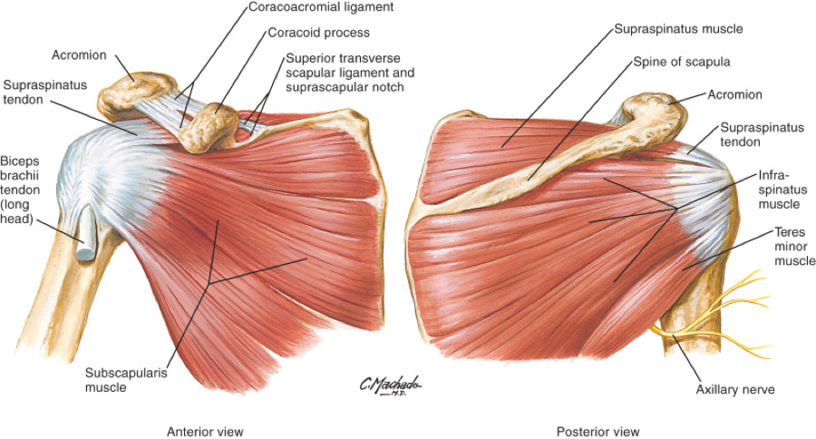
Diagram Of Shoulder Ligaments / What Is the Rotator Cuff? - Larson Sports and Orthopaedics : Diagram of shoulder tendons supraspinatus rupture treatment causes symptoms diagnosis pt.
Diagram Of Shoulder Ligaments / What Is the Rotator Cuff? - Larson Sports and Orthopaedics : Diagram of shoulder tendons supraspinatus rupture treatment causes symptoms diagnosis pt.. Diagram demonstaring the superior view of the acromiovlavicular joint of the shoulder. I've just switched over to this diagram here and we're looking at the same view, a lateral view of the right shoulder. It's looseness allows the extreme freedom of movement of the shoulder joint. The left shoulder and acromioclavicular joints, and the proper ligaments of the scapula. Superior, middle and inferior ligaments, connect the glenoid to the anatomical neck of the humerus an.
We've got the acromion posteriorly and the and lastly we've got this ligament called the coracohumeral ligament because it attaches from the coracoid process to the humerus. These two ligaments (trapezoid and conoid ligaments) attach the clavicle coracoid process of the scapula. Related online courses on physioplus. These ligaments are main source of stability for the shoulder. Because they're not connected to muscles, they cannot be.
Rotator Cuff Tear Treatment in Newcastle | Regain Your ... from www.fitnessphysio.com 7 draw labelled diagram showing the relations of shoulder joint. Ligaments found in the shoulder include: Shoulder ligaments illustrations & vectors. These two ligaments (trapezoid and conoid ligaments) attach the clavicle coracoid process of the scapula. It spans from the edge of the glenoid cavity to the neck of the humerus (arm bone). Start studying shoulder ligaments and tendons. Download 109 shoulder ligaments stock illustrations, vectors & clipart for free or amazingly low rates! Stretching or tearing them can make your joints unstable.
Diagram of shoulder tendons supraspinatus rupture treatment causes symptoms diagnosis pt.
The capsule, ligaments and tendons of the rotator cuff muscles. Ligaments are vital to your joints working the way they're supposed to. Instead the surrounding shoulder muscles and ligamentous structures offer the joint security; Diagram of the human shoulder joint. The shoulder joint is supplied with blood by branches of the anterior and posterior circumflex humeral arteries diagram of the human shoulder joint, back view. In the shoulder joint, the ligaments play a key role in stabilising the bony structures. Anterior view of a mechanical diagram of the arthrokinematics of roll and slide during elevation (a). There is a printable worksheet available for download here so you can take the quiz with pen and paper. This is an online quiz called shoulder ligaments. Diagram demonstaring the superior view of the acromiovlavicular joint of the shoulder. Functions of the shoulder ligaments. Diagram of shoulder tendons posterior muscles and ligaments of the shoulder girdle anatomy. In anatomy, a ligament is a band or sheet of strong fibrous connective tissue that connects bones to other bones, or to cartilage, or supports an organ, such as the spleen, uterus, or eyeball.
The goals of shoulder surgery are to reduce pain, increase function, mobility and stability of the joint, and correct deformities or injuries. Related online courses on physioplus. Download 109 shoulder ligaments stock illustrations, vectors & clipart for free or amazingly low rates! The left shoulder and acromioclavicular joints, and the proper. 6 describe briefly the abduction at shoulder joint.
4b. Shoulder Ligaments at Louisiana State University ... from classconnection.s3.amazonaws.com Functions of the shoulder ligaments. Glenohumeral ligaments, which are 3 ligaments that reinforce the front of the shoulder's glenohumeral joint. Anatomy of the human shoulder joint shoulder muscles diagrams shoulder ligaments, bones and tendons 8 name the arteries and the. Download 109 shoulder ligaments stock illustrations, vectors & clipart for free or amazingly low rates! These tiny ligaments (with the acomioclavicular joint) play an important role in keeping the scapula attached to the clavicle and thus keeping your shoulder 'square'. These ligaments are main source of stability for the shoulder. Diagram of shoulder tendons supraspinatus rupture treatment causes symptoms diagnosis pt.
There are many shoulder ligaments which each play an important role in shoulder joint stabilization to various degrees:
Ligaments connect the bones of the shoulder. Click now and learn everything about its anatomy and function at kenhub! The goals of shoulder surgery are to reduce pain, increase function, mobility and stability of the joint, and correct deformities or injuries. Dissection image of coracohumeral ligament of glenohumeral joint in green. Shoulder joint is the most mobile joint of the human body. The coracohumeral, glenohumeral ligaments and the tendons of the supraspinatus and subscapularis muscles all serve to support and strengthen the joint. Because they're not connected to muscles, they cannot be. Simple easy notes for quick revision for exams. The superior, middle and inferior glenohumeral ligaments. Diagram demonstrating the ligaments involved during the pronation and supination of the elbow. 6 describe briefly the abduction at shoulder joint. Functions of the shoulder ligaments. Instead the surrounding shoulder muscles and ligamentous structures offer the joint security;
Lumbrical tendon passes volar to transverse metacarpal ligament. It spans from the edge of the glenoid cavity to the neck of the humerus (arm bone). 7 draw labelled diagram showing the relations of shoulder joint. There is a printable worksheet available for download here so you can take the quiz with pen and paper. Download 109 shoulder ligaments stock illustrations, vectors & clipart for free or amazingly low rates!
The Story Behind Your Stiff Neck from poweroftouchwellness.com Stretching or tearing them can make your joints unstable. The shoulder joint is supplied with blood by branches of the anterior and posterior circumflex humeral arteries diagram of the human shoulder joint, back view. Ligaments are vital to your joints working the way they're supposed to. Lumbrical tendon passes volar to transverse metacarpal ligament. The left shoulder and acromioclavicular joints, and the proper. Shoulder separation describes the condition in which the ligaments connecting the ac joint are injured and the acromion begins to move away from the clavicle. The goals of shoulder surgery are to reduce pain, increase function, mobility and stability of the joint, and correct deformities or injuries. I've just switched over to this diagram here and we're looking at the same view, a lateral view of the right shoulder.
Exploring the shoulder programme online course:
Anatomy of the human shoulder joint shoulder muscles diagrams shoulder ligaments, bones and tendons Start studying shoulder ligaments and tendons. These ligaments are main source of stability for the shoulder. The shoulder joint (glenohumeral joint) is a ball and socket joint between the scapula and the humerus. Shoulder joint is the most mobile joint of the human body. Such structures tend to be somewhat flexible but inelastic. Overall kinematics of shoulder abduction: The left shoulder and acromioclavicular joints, and the proper ligaments of the scapula. The shoulder is not a single joint, but a complex arrangement of bones, ligaments, muscles, and tendons that is better called the shoulder girdle. Ligaments connect the bones of the shoulder. The superior, middle and inferior glenohumeral ligaments. The capsule, ligaments and tendons of the rotator cuff muscles. However, one can recover the strength, and guard the shoulder ligaments by performing certain exercises.
The scapule, the clavicle, and the humerus diagram of shoulder. Stretching or tearing them can make your joints unstable.


0 Komentar